Deep brain stimulation

Deep Brain Stimulation (DBS)

More than 150,000 patients have undergone DBS. The present day techniques make it a very safe procedure.

Surgical Procedure of DBS

Various components of the DBS system need to understood before discussing the surgical steps in details. Though, as a patient, you may not need to get to choose between them, your doctor can best advise you on the most appropriate solution.

Components of DBS ( Deep Brain Stimulation) system

DBS implant system comprises of 3 parts: The lead, Internal Pulse Generator (IPG)& the extension wire.

The lead: This is a thin coiled wire insulated in polyurethane with four platinum iridium electrodes at its tip.

The Internal Pulse Generator (IPG): The IPG is a battery-powered neurostimulator housed in a titanium casing, that sends electrical pulses to the brain to interfere with neural activity by neurons at the target site.

The Extension wire: It’s an insulated wire. The lead is connected extra-cranially to the IPG by the extension wire.

Placement of different components of DBS System:

The placement of lead is done in the deep targeted region of the brain through a small burr hole with extracranial end coming out of it through the burr hole.

The extension wire connects to the extracranial end of the leads to the IPG& it runs from the head, down the side of the neck, behind the ear to the IPG.

The IPG is placed in a subcutaneous pouch about 1inch below the clavicle over the left anterior chest wall.

Selection of patients:

Jaslok Hospital & Research Centre, Mumbai has got a sophisticated and world class state of art Functional Neurosurgery department for surgical treatment for Parkinson’s disease & other movement disorders.

This department is being headed by Dr. Paresh Doshi, a famous well-known skilled Functional & Stereotactic Neurosurgeon. Patients were consulted in his out-patient department& appropriate patients are selected for surgery.

Parkinson’s disease patients who had responded well to levodopa therapy early in the course of their disease but presented with severe motor fluctuations to medications with increased time period of ‘OFF phase’ in a day are the right candidates chosen for deep brain stimulation.

After evaluation, the selected candidates are explained about the risks and benefits out of DBS surgery for Parkinson’s disease. Video footage of previous successful patients are displayed among some apprehensive patients and their relatives to allay their fear about the surgery.

Preparation for Surgery & surgical process:

The selected patients for surgery are instructed to not to take any drugs like aspirin, clopidogrel or any blood thinner 10 days prior to Deep Brain Stimulation surgery, as these drugs can increase the risk of bleeding during the surgery.

Patient should be free from any kind of infection e.g. urinary tract infection or dental infection because presence of infection can spread to implant sites leading to implant failure. Therefore, surgical team should be made aware of the presence of infection prior to surgery.

Steriotactic Biopsy -surgical-procedures

Approximately 10 to 12 days of hospitalisation of the patient is needed for Deep Brain Stimulation surgery. History & neurological examination of the patient is done on admission by a resident doctor.

Any morbid medical conditions such as heart disease, Hypertension and diabetes etc. have to be ruled out & the patients are advised to disclose about any such illness for better evaluation prior to the surgery.

Routine investigations such as blood test, ECG, 2D Echo, X-Ray of chest as well as pre anaesthetic check-up are also carried out on the patient prior to the surgery.

The patient has to undergo Unified Parkinson’s Disease Rating Scale (UPDRS) examination in OFF and ON medications for evaluation of the severity of Parkinson’s disease.

Mini-mental status examination (MMSE)to evaluate memory & judgement issues. For OFF phase UPDRS examination, all anti Parkinson’s medications are stopped at around 9pm on the day of admission as almost 12 hrs are required to ward off the effects of anti-parkinsonian drugs.

For ON phase evaluation, the patient is given 2 times his usual dose of L-dopa and the UPDRS is repeated. Video recording of patient’s motor activity is done in both OFF & ON phase.

Patient’s caretakers are supplied with Parkinson’s Disease Questionnaire 39 Items (Global QoL) and Zerith Caretaker Burden Inventory (ZCBI) which has to be filled by them.

Pre-operative 3 Tesla Magnetic Resonance Image (MRI) of the brain is done in DBS protocol that serves as a roadmap during DBS surgery for Parkinson’s disease by the neurosurgeon to ensure proper placement of electrodes.

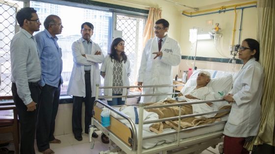
Parkinson’s disease nurse helps in promoting a relaxed and supportive environment in pre-operative waiting period as well as during surgery. Patient cooperation and comfort during the DBS surgery is highly essential & patients are encouraged to express their needs so as ensure a relaxed ambience.

PD nurse spends considerable time explaining the surgical procedures with the patient and their relatives to relieve their anxiety. Repeated interaction with the patient boosts confidence in him regarding surgery.

Sometimes this conversation with the patient brings out important observations by the PD nurse that helps the surgeon & his surgical team conducting a safe and smooth surgery on the patient.

Personalized care from a well-informed team of nurses & paramedics within the ward allow the patients to gain confidence over the caretakers during the stay in the hospital.

The pre-op preparation starts with pre-anaesthetic check up on the 2nd day evening by the anesthesiologist who thoroughly evaluate patient’s medical fitness prior to surgery.

This is followed by head shaving, after obtaining due consent. Consent for surgery should also be obtained from the patients and their spouse or close relatives. Anti-Parkinson’s medications are stopped around 10 pm & the patient should be kept in empty stomach.

At around 4 A.M. patient is given a banana and a glass of milk or a chocolate to prevent vasovagal syncope during surgery.

Patient should be bathed properly and kept ready at 6 A.M. to be shifted to operation theatre for surgery. Once everything is ready, the resident doctor shifts the patient to Operation theater at around 7.30 A.M.

The OT staffs begin preparation for surgery that includes insertion of an IV canula, administration of antibiotics & IV fluids as well as monitoring of the patient’s vital signs like Blood pressure & pulse. Urinary catheter is inserted by the resident doctor after shifting the patient to OT table.

The duration of entire surgical process is around 5 to 6 hours. The placement of stereotactic frame is done under local anaesthesia. It consists of ahead ring which is attached at 4 points (two anteriorly & two posteriorly) to the patient’s skull.

The placement of the frame is done under monitored anaesthesia care under direct supervision of an anaesthesiologist with minimal sedation, to ensure patient comfort.

The procedure is done under local anaesthesia & the patient remains awake throughout the surgical procedure. Surgery starts with drilling of burr holes on the top of the skull at desired points predefined by functional planning.

As both bone & brain are insensitive to pain, this surgery becomes a relatively painless procedure by infiltration of local anaesthesia into the scalp. After burr holes are made, thin microelectrodes are inserted into the predefined targets deep inside the brain & physiological brain mapping is done by a procedure called micro-electrode recordings.

Once the exact location is identified by MER, a low voltage current (micro stimulation) is administered through the macroelectrodes to find out the level of improvement that can be achieved by DBS.

This is followed by checking of side effects by increasing the micro stimulation. Patient’s cooperation is highly necessary at this stage.

The level of improvement is judged by arrest of tremors in patients experiencing tremors, relief from pain in patients presenting with pain and there will be decrease in stiffness in patients presenting with Parkinson’s disease on stimulating the correct target site.

Permanent DBS lead is implanted under fluoroscopic guidance once adequate confirmation of the target site is obtained by the surgical team though MER mapping & response to micro stimulation.

Post-operative procedures

Following DBS surgery, a check CT scan of brain is performed for confirmation of the position of electrodes and rule out any complications like intracranial hematoma or tension pneumocephalus (trapping of air inside intracranial cavity).

After the CT scan is over, the patient is transferred either to the ward or the ICU for observation depending upon his condition. Here he is administered the scheduled dose of his L-dopa and allowed to have his meals afterwards.

Deep-brain-stimulation-surgery-process-Paresh Doshi
Deep-brain-stimulation-dr.-paresh-doshi

Usually on the next day morning, a pace maker or battery also called as IPG (Implantable Pulse Generator) is implanted 2 inches below the left clavicle on the chest wall by creating a subcutaneous pouch under general anesthesia.

Just after surgery, most of the patients experience improvement in the symptoms. Thesis usually due to be micro lesioning effect caused by the actual electrode placement in the brain and the swelling of the brain after the lead placement.

After the lesioning phase is over, the IPG is activated and the programming is done to see the response of the lead contact points in the form of improvements in disease symptoms and also their side effects.

Good programming involves appropriate settings on good contact points with reduction in the doses of anti-Parkinson’s medications. Once optimum stimulation has been set, the PD Nurse starts educating patients and their family members regarding usage of the remote device supplied by the company.

They were also taught about remote device handling &post-op care of the patient at home. Once the scalp & chest stitches are removed and the dressing is done on the 8th post-op day, patient is discharged Patient is sent back home with instruction for removal of the dressing after two days.

Any bleeding from the wound site or appearance of infection near the wound site should be immediately reported to the surgical team. The patient and their family members are supplied with a list of precautions &an instruction manual of care to be followed for future referral.

The surgical team provides on-going support for the patient& their relatives as well as professionals telephonically.

A discharge summary with detailed procedure followed is provided to the patient, including contact details of the surgical team to be contacted in emergency situation.

The patient’s 1stfollow-up appointment is usually scheduled after three months. Usually four to six months are necessary for stabilization post operatively & active support from surgical team is provided throughout this period.

The goal of this therapy is to achieve symptoms control adequately and effectively with optimum level of stimulation in combination with drug therapy without their side effects.

Patient can adjust the current with the remote device according to their requirement. Routine follow up is usuall