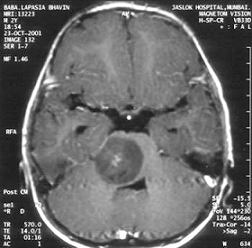
We commonly perform Stereotactic brain Biopsies under local anesthesia with or without any intravenous sedation. Most of the biopsies are performed with the help of CT localization, however, in cases where the lesion cannot be seen on CT, MRI guided biopsies can be done.
The procedure usally takes about 3 hours.
It begins with a stereotactic frame attached to the patient’s head using local anesthesia at the pin insertion points.
This works as a reference point for all the scans (CT, MRI and PET) which are used for target localization. The system allows computerized planning of the surgical approach of the lesion with sub-millimeter precision. A CT/MRI scan is then performed to obtain the co-ordinates to the target.
In the operating room, the patient’s head is to be rested on a clamp system in a comfortable position. A small linear incision is then made on the scalp and a small burhole is drilled into the skull.
A reedy biopsy needle is then put into the brain using the coordinates obtained by the computer workstation to the target lesion. This is a very less invasive and much more precise than an open biopsy procedure that requires a craniotomy which involves removing of a piece of the skull in order to get access to the brain lesion.
The specimen is then sent to the pathologist for histopathologic evaluation who will give opinion, if the tissue is representative of the lesion targetted and adequate for the final diagnosis.
Post-operatively, a CT scan is done to rule out any haemorrhage and also to confirm the site of biopsy taken.
The patient is then kept under observation following the procedure and usually goes to home within 1-2 days.